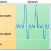
Breathing (inspiration and expiration) occurs in a cyclical manner due to the movements of the chest wall and the lungs. The resulting changes in pressure, causes changes in lung volumes, i.e. the amount of air the lungs are capable of occupying. These volumes tend to vary, depending on the depth of respiration, ethnicity, gender, age and in certain respiratory diseases.
Tidal Volume (TV)
The volume of air breathed in and out at rest is known as the tidal volume (TV). This is found to be about 500 ml in an averagely built (70 kg), healthy, young adult. The tidal volume tends to decrease in restrictive lung diseases. In restrictive lung diseases, the lungs fail to expand properly as a result of restrictive forces exerted from within the lungs (e.g. – fibrosing alveolitis) or from the thoracic wall (e.g. – severe scoliosis, ankylosing spondylitis). Weakness of the respiratory muscles (e.g. – myasthenia gravis, Guillain Barre syndrome and phrenic nerve palsy) can also give rise to restricted movements of the chest wall resulting in the reduction of the tidal volume.
Inspiratory Reserve Volume (IRV) and Expiratory Reserve Volume (ERV)
In addition to the amount of air that could be inspired at rest, the lungs are capable of accommodating an additional amount of air during a deep inspiration. This amount of air that can be inhaled in addition to the tidal volume is known as the inspiratory reserve volume (IRV). Similarly, in a deep and forceful expiration, the lungs are capable of exhaling a volume which is in excess to the tidal volume and the inspiratory reserve volume. This is known as the expiratory reserve volume (ERV). In a healthy young adult, IRV measures about 3 L and the ERV is approximately 1.3 – 1.5 L.
Residual Volume (RV)
The lungs do not collapse completely following a deep, forceful expiration. A certain volume of air remains within the lungs, maintaining the alveoli expanded and the airways patent. This volume, which cannot be expelled even after a maximally forceful expiration, is known as the residual volume (RV).
Measurement of Lung Volumes – Spirometry
The tidal volume, IRV and ERV can be measured using a device known as a spirometer. Here, the volume changes that occur in a closed circuit are measured while an individual is breathing through a mouth piece into a measuring device. The volume change that occurs while the individual is engaged in quite breathing is the tidal volume. The volume that the individual inhales in excess of the tidal volume during a deep inspiration is the IRV and the volume that is exhaled in excess to the tidal volume during a deep expiration is the ERV.
Measurement of the Residual Volume – Helium Dilution
The residual volume cannot be measured with a conventional spirometer. Therefore, to measure the residual volume, several techniques have been described. In one such technique, an individual breaths into a closed circuit, which contains a known amount of Helium. Helium does not cross the blood-gas barrier and is not excreted by the lungs. Thus, decrease in the concentration of Helium is brought about by the increase in the volume of the circuit by connecting the circuit to the respiratory system. When, the concentration of Helium is measured following a deep expiration, the total volume is the volume of the breathing circuit + the residual volume. Since concentration = amount of a substance / volume of distribution, the residual volume can be calculated.
Lung Capacities………
Four capacities have been described based on the four lung volumes:
Inspiratory Capacity (IC) is the maximum volume of air that can be inhaled following a resting state. This can be calculated by the addition of tidal volume and the IRV.
Vital Capacity (VC) is the maximum volume of air that can be exhaled following a deep inspiration. This is the total of IRV + TV + ERV.
Functional Residual Capacity (FRC) is the volume of air that remains in the lungs during quite breathing. FRC = ERV + RV.
Total Lung Capacity (TLC) is the volume the whole respiratory system can accommodate. Therefore, TLC= IRV + TV + ERV + RV.
Lung capacities and lung volumes are affected in different types of physiological processes as well as in lung diseases. The specific changes that occur in different types of diseases will be described in a separate hub along with examples for different patterns of abnormalities seen in the lung volumes.
, Lung Volumes and Capacities www.ozeldersin.com bitirme tezi,ödev,proje dönem ödevi
